


ELECTRODIAGNOSTICS
BAER TEST
Brainstem auditory evoked response
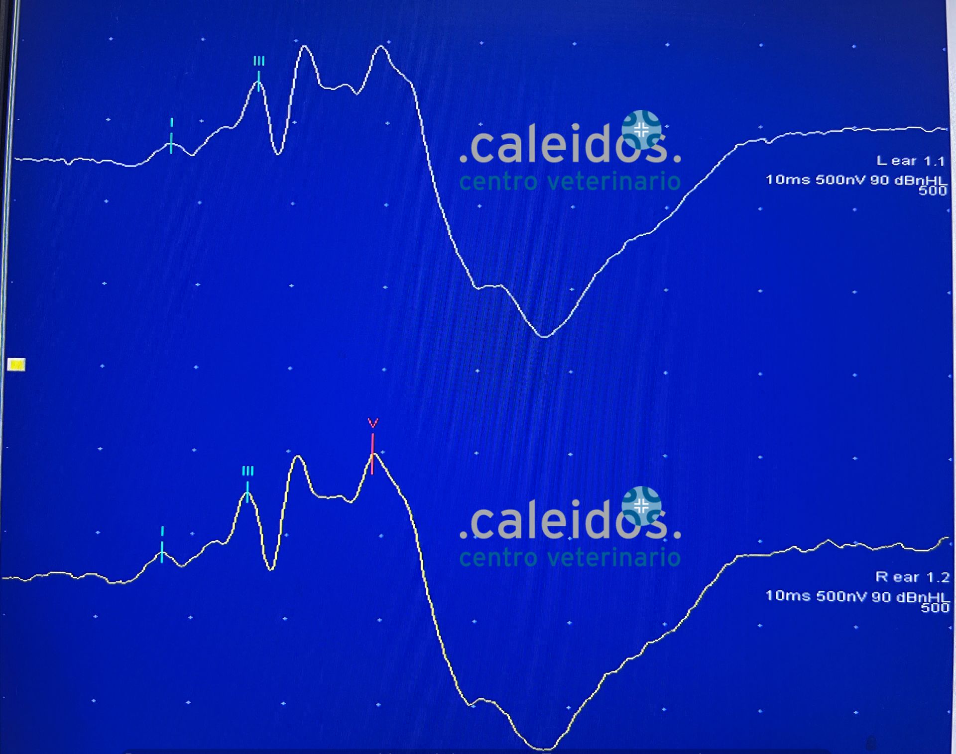
The BAER test allows you to objectively identify the deafness of an animal, using small electrodes connected to a device that sends sound impulses and records the brain's response to them. The latter will then be converted into a path, made up of waves of different amplitudes and heights.
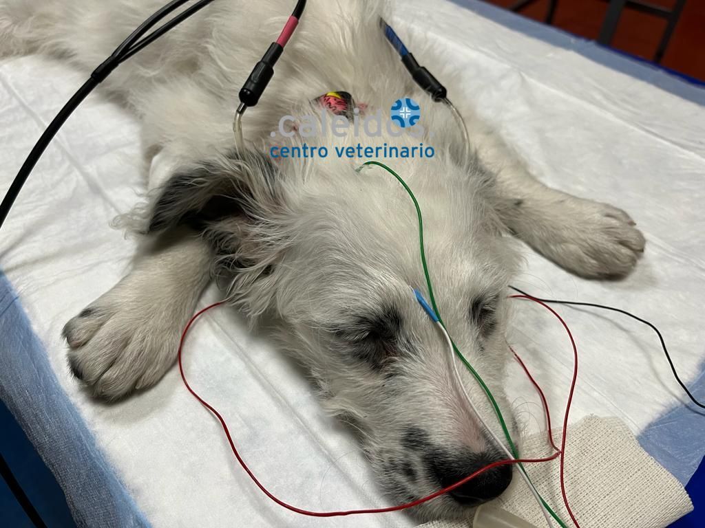
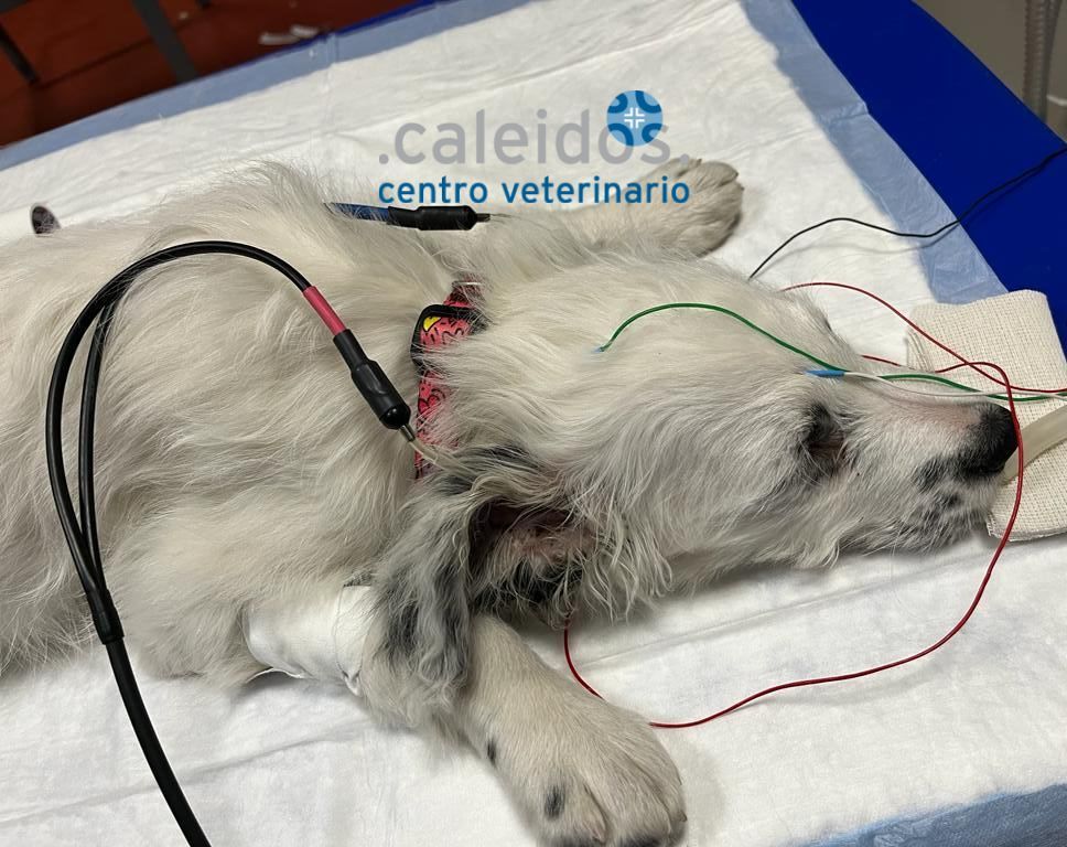
Often the test is performed by subjecting the dog or cat to mild sedation so that its movements do not interfere with the outcome; since deafness can be in one ear or both, one ear is analysed at a time.
BAER Test purposes:
- To identify forms of congenital deafness affecting puppies of genetically predisposed breeds, in which early degeneration of the cochlea, the inner ear structure responsible for converting acoustic information into nerve impulses, may occur.
Approximately eighty breeds have been identified with a hereditary form of deafness, with Dalmatians, Bull Terriers, English Setters, Cocker Spaniels, and other dogs with white coats being the most affected, especially when combined with light-colored eyes.
It has been observed that dogs with congenital deafness can be trained to respond to signals other than acoustics, such as hand movements.
- To diagnose late-onset forms secondary to other conditions. In such cases, MRI is used, enabling the differentiation of conditions affecting the middle ear, inner ear, and brain structures associated with hearing loss.
Deafness linked to otitis externa, if properly treated, will be reversible and the BAER test will show the recovery of hearing function.
ELECTROMYOGRAPHY
An electromyographic examination provides data that allow us to distinguish between a muscle disease (myopathy, dystrophies, myositis, myasthenia) and a disease of the peripheral nervous system (polyneuropathy, neuritis, nerve compression syndrome in a peripheral nerve root), making it a useful support in diagnosing and classifying many neurological diseases.
Electromyography (EMG) is the recording of muscle electrical activity. In a healthy subject, when the muscle is at rest, no electrical activity is recorded, while during a voluntary contraction, gradually more intense up to the maximal, progressive recruitment of the motor unit potentials (MUPs) is observed, until the "interference" framework is reach, in which the individual MUPs are no longer distinguishable from each other.
Although the electromyographic study cannot provide data on the aetiology of the injuries, it can be used to better define their location, extension and extent and can also be useful for monitoring the evolution of a pathology.


